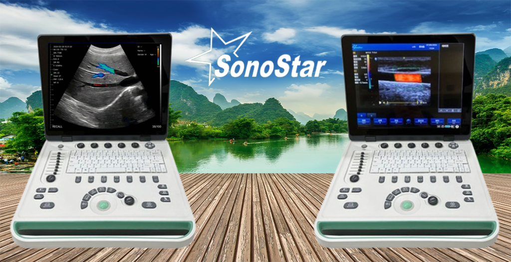
Portable Color Doppler Ultrasound Machine

US

I’m Looking For
Why Every Physician Should Have a Portable Ultrasound Machine
A portable ultrasound machine can be a game-changer for physicians in various specialties. Here’s why every physician might benefit from having one and how to choose the best option:
Enhanced Diagnostic Capability
- Provides real-time imaging to aid in diagnosis.
- Reduces dependency on external imaging facilities.
Point-of-Care Use
- Ideal for bedside evaluations in clinics, hospitals, or even home visits.
- Speeds up decision-making for critical conditions.
Improved Patient Outcomes
- Early and accurate diagnosis can lead to better treatment plans.
- Minimizes the need for invasive procedures.
Versatility Across Specialties
1. General Medicine
- Point-of-Care Ultrasound (POCUS): Quick bedside assessments of internal organs, fluid collections, or abscesses.
2. Emergency Medicine
- FAST Exam: Focused Assessment with Sonography for Trauma to detect internal bleeding.
- Cardiac Arrest: Evaluate cardiac activity during resuscitation.
3. Cardiology
- Echocardiography: Assess heart function, detect valve issues, and measure ejection fraction.
- Vascular Imaging: Identify deep vein thrombosis (DVT) or arterial blockages.
4. Obstetrics and Gynecology
- Fetal Monitoring: Check fetal growth, position, and amniotic fluid levels.
- Pelvic Imaging: Diagnose conditions like ovarian cysts, ectopic pregnancy, or uterine abnormalities.
5. Anesthesiology
- Nerve Blocks: Guide needle placement for regional anesthesia.
- Vascular Access: Assist in placing central or peripheral lines.
6. Critical Care
- Lung Ultrasound: Detect pneumothorax, pleural effusion, or pulmonary edema.
- Cardiac Output Monitoring: Assess fluid responsiveness in critically ill patients.
7. Musculoskeletal (MSK) Medicine
- Joint and Tendon Imaging: Diagnose injuries like rotator cuff tears or arthritis.
- Fracture Assessment: Evaluate bone integrity in suspected fractures.
8. Sports Medicine
- Soft Tissue Injuries: Identify sprains, strains, or muscle tears.
- Guided Injections: Accurately place corticosteroid or PRP injections.
9. Rheumatology
- Joint Inflammation: Detect and monitor synovitis or effusion in patients with arthritis.
10. Urology
- Bladder Scanning: Measure post-void residual volume.
- Kidney Imaging: Detect stones, hydronephrosis, or masses.
11. Gastroenterology
- Abdominal Imaging: Evaluate liver, gallbladder, pancreas, and spleen.
- Ascites Detection: Identify and monitor fluid accumulation in the abdomen.
12. Pediatrics
- Neonatal Brain Ultrasound: Assess brain structures in premature infants.
- Pediatric Lung Imaging: Diagnose pneumonia or pleural effusions in children.
13. Pulmonology
- Thoracic Imaging: Assess pleural effusions, pneumothorax, or consolidation.
- Diaphragm Function: Evaluate diaphragmatic movement in respiratory failure.
14. Endocrinology
- Thyroid Imaging: Detect nodules, cysts, or enlargement.
- Parathyroid Scans: Identify abnormal parathyroid glands.
15. Oncology
- Tumor Detection: Locate and assess tumors in the abdomen, breast, or thyroid.
- Guided Biopsies: Assist in taking tissue samples from suspicious masses.
16. Dermatology
- Soft Tissue Masses: Evaluate skin lesions, cysts, or lipomas.
- Vascular Lesions: Assess blood flow in hemangiomas.
17. Ophthalmology
- Orbital Ultrasound: Examine the eye for retinal detachment or tumors.
- Optic Nerve Measurement: Assess for increased intracranial pressure.
18. Podiatry
- Foot and Ankle Imaging: Diagnose plantar fasciitis or Achilles tendon injuries.
- Vascular Assessment: Monitor blood flow in patients with diabetes.
19. Veterinary Medicine
- Animal Diagnostics: Evaluate internal organs, pregnancies, or injuries in pets or livestock.
20. Preventive Care
- Health Screenings: Early detection of vascular disease, liver fibrosis, or abdominal aortic aneurysm in routine checkups.
21. Breast Surgery
1. Preoperative Assessment
- Tumor Localization: Ultrasound is used to identify the exact size, location, and extent of breast tumors, especially for non-palpable lesions. This helps in planning the surgical approach (e.g., lumpectomy or mastectomy).
- Axillary Lymph Node Evaluation: Ultrasound helps detect lymph node involvement, guiding decisions about sentinel lymph node biopsy or axillary dissection.
2. Intraoperative Guidance
- Real-Time Navigation: During surgery, ultrasound provides real-time imaging to ensure complete tumor removal, minimizing residual disease.
- Margin Assessment: Helps surgeons evaluate surgical margins to confirm clear resection, reducing the likelihood of reoperation.
