Optimizing Accuracy and Patient Safety in Vascular Access with Handheld Wireless Color Doppler Ultrasound Technology
Have you ever struggled with achieving accurate vascular access, especially in high-pressure or emergency scenarios? Can you recall instances where conventional techniques failed to offer sufficient guidance for vein cannulation, resulting in treatment delays or complications? Picture a situation where precise vessel identification is critical, yet traditional tools fall short. How frequently do healthcare providers face obstacles in securing vascular access in patients with complex anatomies, or in cases involving pediatric, elderly, or obese patients?
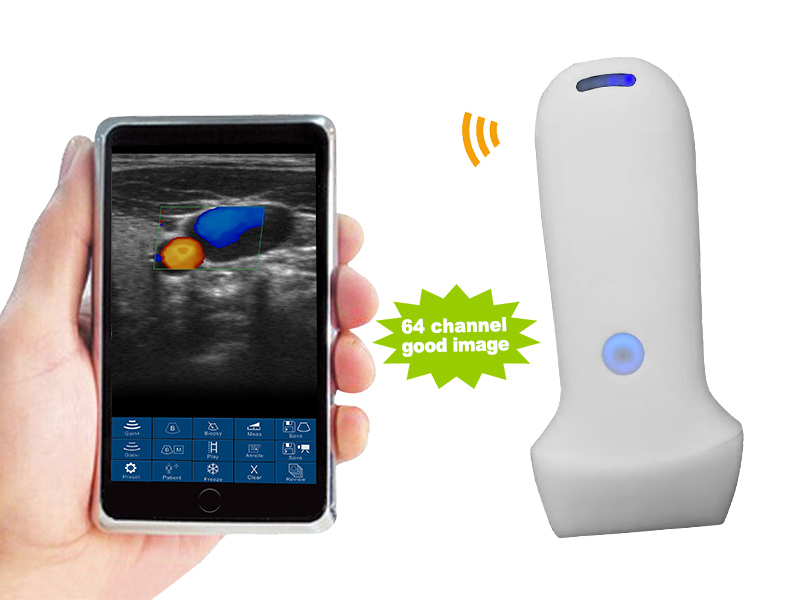
Suitable Handheld Wireless Color Doppler Ultrasound for Vascular Medicine
Linear array Frequency: 7.5MHz/10.0MHz


- Linear modes.
- Comprehensive connectivity with support for iOS, Android, and Windows platforms.
- Frequency: Linear probe 7.5MHz/10.0MHz
- Display mode: B, B/M, color doppler version with B+Color, B+PDI, B+PW
- Display Depth: Linear 20/40/60/80mm
- Measure: Length, Area, Velocity, HR, S/D, Auto Blood Vessel Measurement
- Weight: 180g
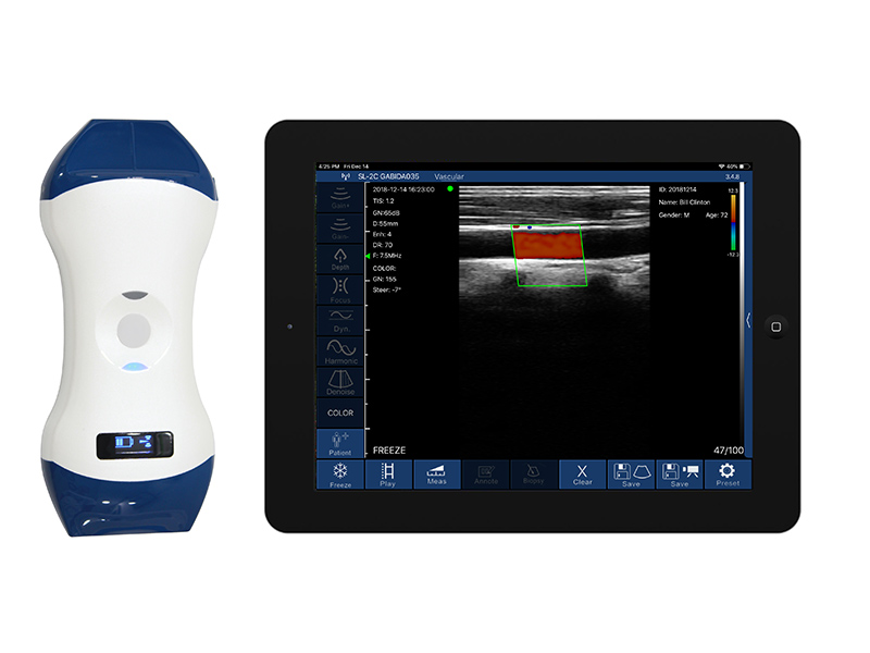
Double Headed Handheld Wireless Color Doppler Ultrasound for Physical Medicine
Linear array Frequency: 7.5MHz/10.0MHz & 10MHz/12.0MHz


- Linear modes.
- Comprehensive connectivity with support for iOS, Android, and Windows platforms.
- Frequency: Linear probe 7.5MHz/10.0MHz & 10MHz/12.0MHz
- Display mode: B, B/M, color doppler version with B+Color, B+PDI, B+PW
- Display Depth: Linear 20/40/60/80mm
- Measure: Length, Area, Velocity, HR, S/D, Auto Blood Vessel Measurement
- Weight: 250g
Handheld Wireless Color Doppler Ultrasound: Key Applications for Vascular Access
Handheld wireless color Doppler ultrasound technology has become an indispensable tool for vascular access, providing real-time guidance, enhancing accuracy, and minimizing complications during procedures. Its portability, high-resolution imaging, and Doppler capabilities make it especially useful for evaluating and accessing veins and arteries in various clinical settings.
Here are the key applications for handheld wireless color Doppler ultrasound in vascular access:
1. Peripheral Venous Access
- Application: Facilitating the insertion of peripheral IV lines, particularly in patients with difficult venous access (DVA) due to conditions like obesity, dehydration, or chronic illness.
- Details:
- Real-Time Visualization: Ultrasound enables clinicians to visualize veins in real time, ensuring accurate catheter placement, even in challenging cases where veins are not visible or palpable.
- Color Doppler: Color Doppler helps assess blood flow within the vein, confirming the vessel’s patency and avoiding inadvertent puncture of arteries or collapsed veins.
- Minimizing Complications: Using ultrasound guidance reduces complications like hematoma, inadvertent arterial puncture, and multiple needle sticks.
2. Central Venous Catheterization (CVC)
- Application: Ultrasound guidance for the placement of central venous catheters (CVC) in the internal jugular, subclavian, or femoral veins.
- Details:
- Improved Accuracy: Ultrasound-guided CVC placement ensures that the needle is directed into the vein, avoiding complications like pneumothorax and arterial puncture.
- Doppler for Vessel Differentiation: Color Doppler helps distinguish between arteries and veins, reducing the risk of arterial cannulation.
- Real-Time Monitoring: Clinicians can continuously monitor the needle’s trajectory and confirm proper catheter placement, reducing the risk of catheter misplacement or vessel injury.
3. Arterial Access for Monitoring and Blood Gas Analysis
- Application: Assisting in the insertion of arterial lines for continuous monitoring of blood pressure or for arterial blood gas (ABG) sampling, often required in critically ill patients.
- Details:
- Precise Identification: Color Doppler ultrasound aids in identifying arteries, such as the radial, femoral, or brachial artery, ensuring precise needle placement.
- Real-Time Visualization: The ability to visualize the artery and surrounding structures in real time reduces the likelihood of complications such as arterial dissection or hematoma formation.
- Minimized Failure Rates: Ultrasound guidance significantly increases the success rate of first-attempt arterial cannulations, especially in patients with compromised circulation or hypotension.
4. Peripherally Inserted Central Catheter (PICC) Lines
- Application: Guiding the placement of PICC lines in patients requiring long-term intravenous access for antibiotics, chemotherapy, or total parenteral nutrition (TPN).
- Details:
- Vein Selection: Ultrasound helps identify suitable veins, such as the basilic or cephalic veins, ensuring that a vein with adequate diameter is selected to accommodate the PICC line.
- Doppler Imaging: Color Doppler can confirm vein patency and detect any venous thrombosis or narrowing that could complicate PICC placement.
- Reduced Complications: By using real-time ultrasound guidance, clinicians can avoid multiple needle insertions, minimizing the risk of complications like infection and vein trauma.
5. Dialysis Catheter Placement
- pplication: Assisting in the placement of dialysis catheters in patients with end-stage renal disease (ESRD) who require hemodialysis.
- Details:
- Identifying Suitable Veins: Ultrasound is used to locate and assess central veins (e.g., internal jugular or subclavian veins) and peripheral veins for temporary dialysis access.
- Color Doppler for Blood Flow: Color Doppler imaging confirms adequate blood flow and ensures that the selected vein is patent for dialysis catheter insertion.
- Prevention of Complications: Ultrasound-guided dialysis catheter placement reduces the risk of vascular injury, pneumothorax, and misplacement of the catheter.
6. Vascular Access in Pediatric Patients
- Application: Assisting with vascular access in pediatric patients, where veins are smaller and more difficult to visualize or palpate.
- Details:
- Minimizing Trauma: Handheld ultrasound provides a non-invasive method for visualizing pediatric veins, reducing the number of attempts and minimizing trauma for children during venous or arterial access.
- Doppler for Vessel Patency: Doppler ultrasound ensures the chosen vessel is patent and has adequate blood flow, critical for the smaller anatomy of pediatric patients.
- Increased Success Rates: Ultrasound guidance is particularly valuable in neonates or infants, where vascular access can be extremely challenging without visualization.
7. Difficult Intravenous Access (DIVA) in Adult and Geriatric Patients
- Application: Ultrasound-guided vascular access for patients with difficult intravenous access (DIVA), often encountered in elderly patients, patients with chronic illnesses, or IV drug users.
- Details:
- Visualizing Deep Veins: Ultrasound enables visualization of deep veins that may not be palpable or visible, increasing the success rate of IV access in geriatric patients with fragile veins.
- Doppler for Blood Flow: Doppler imaging ensures that veins selected for cannulation are not collapsed or thrombosed, which is common in patients with multiple comorbidities or a history of frequent venous access.
8. Assessment of Vascular Complications
- Application: Evaluating patients for vascular complications following central or peripheral venous catheterization, including thrombosis, hematoma, or arterial injury.
- Details:
- Thrombosis Detection: Color Doppler ultrasound can detect deep vein thrombosis (DVT) or other clots that may occur as a complication of vascular access, enabling early intervention.
- Assessing Hematomas: Ultrasound imaging helps assess the extent of hematomas around insertion sites and guide the management of complications like arterial puncture or extravasation.
- Monitoring Post-Procedure: Ultrasound allows for regular monitoring of the vascular access site, ensuring that the catheter remains patent and without complications.
9. Vascular Access for Outpatient Procedures
- Application: Assisting with vascular access in outpatient procedures requiring intravenous access, such as sedation, contrast imaging, or infusion therapy.
- Details:
- Time Efficiency: Handheld ultrasound enables rapid, accurate placement of IV lines in outpatient settings, improving the patient experience and reducing delays in initiating treatments.
- Reducing Procedure Time: By providing clear visualization of veins and arteries, ultrasound reduces the time needed for venous cannulation, especially in patients with difficult venous access.
10. Assessment of Arteriovenous (AV) Fistulas and Grafts
- Application: Evaluating and monitoring arteriovenous (AV) fistulas or grafts in patients with chronic kidney disease undergoing hemodialysis.
- Details:
- Patency Assessment: Color Doppler ultrasound allows for real-time evaluation of blood flow through AV fistulas or grafts, ensuring they are functioning properly for dialysis.
- Detection of Stenosis or Thrombosis: Doppler imaging can detect narrowing or clot formation within the fistula or graft, enabling early intervention to prevent dialysis failure.
- Guiding Interventions: Ultrasound can guide procedures such as angioplasty or thrombectomy for AV fistulas and grafts, improving treatment outcomes.
11. Post-Surgical Vascular Monitoring
- Application: Monitoring vascular grafts and arterial bypasses after surgery to ensure proper function and blood flow.
- Details:
- Doppler for Flow Evaluation: Post-surgical ultrasound is used to assess the blood flow through grafts and bypasses, ensuring that they remain patent and function properly.
- Early Detection of Complications: Doppler can detect early signs of thrombosis or stenosis, allowing for timely interventions to prevent graft failure.
12. Vascular Malformation Diagnosis
- Application: Diagnosing and assessing vascular malformations such as arteriovenous malformations (AVMs) or hemangiomas.
- Details:
- Flow Patterns: Doppler ultrasound evaluates abnormal blood flow patterns within the malformation, helping to differentiate between low-flow and high-flow lesions.
- Real-Time Monitoring: Color Doppler can monitor the progression of malformations over time, aiding in treatment planning and follow-up care.
13. Vascular Injury or Trauma Assessment
- Application: Evaluating patients with suspected vascular injuries or trauma to blood vessels, especially after accidents or surgeries.
- Details:
- Immediate Visualization: Handheld ultrasound provides rapid imaging to detect arterial or venous tears, hematomas, or pseudoaneurysms in trauma settings.
- Doppler Imaging: Real-time color Doppler helps assess blood flow in damaged vessels, allowing clinicians to determine the extent of injury and guide treatment.
14. Peripheral Arterial Disease (PAD) Evaluation
- Application: Assessing patients with suspected peripheral arterial disease by visualizing blood flow in lower extremity arteries.
- Details:
- Color Doppler Imaging: Used to assess the patency of arteries and detect areas of stenosis or occlusion.
- Waveform Analysis: Doppler can evaluate flow patterns to distinguish between normal and obstructed arteries, helping clinicians determine the severity of the disease.
15. Carotid Artery Stenosis Evaluation
- Application: Assessing the carotid arteries for signs of narrowing (stenosis) or plaque buildup that could increase the risk of stroke.
- Details:
- Real-Time Imaging: Color Doppler visualizes the carotid arteries and evaluates the intimal thickness and any atherosclerotic plaques.
- Flow Velocity Measurements: Doppler measures the velocity of blood flow to identify high-risk stenosis that may require medical intervention.
16. Venous Insufficiency and Varicose Veins
- Application: Diagnosing chronic venous insufficiency and varicose veins by assessing venous flow and valve function.
- Details:
- Valve Assessment: Doppler ultrasound evaluates the competency of venous valves to diagnose reflux in the great saphenous vein or other superficial veins.
- Flow Patterns: Color Doppler provides detailed visualization of blood flow, helping identify venous insufficiency, which leads to the development of varicose veins.
Handheld wireless color Doppler ultrasound is a powerful tool for vascular imaging, providing real-time insights into blood vessel health, blood flow, and potential pathologies. Its portability, ease of use, and advanced imaging capabilities make it suitable for a wide range of vascular applications, from diagnosing conditions like PAD and DVT to assisting with procedures such as dialysis catheter placement and vascular trauma evaluation. The technology not only enhances diagnostic accuracy but also improves patient outcomes by facilitating early intervention and better procedural guidance in vascular care.
Choosing the ideal Handheld Wireless Color Doppler Ultrasound for Vascular Physicians: Key Factors to Consider
Selecting the right handheld ultrasound device for vascular physicians is crucial for enhancing diagnostic accuracy and improving patient outcomes. Vascular specialists rely on ultrasound technology for a wide range of applications, from assessing blood flow to detecting vascular abnormalities. When choosing a handheld ultrasound for vascular practice, here are key factors to consider:
1. Imaging Quality and Resolution
- Why It Matters: Vascular assessments require high-resolution images to visualize small vessels, detect blockages, and evaluate blood flow accurately.
- What to Look For: Choose a device with excellent B-mode resolution for clear structural imaging and color Doppler sensitivity for detailed blood flow visualization. This ensures clear images for assessing conditions such as deep vein thrombosis (DVT) or arterial stenosis.
2. Color Doppler and Spectral Doppler Capabilities
- Why It Matters: Vascular diagnostics often depend on precise measurements of blood flow velocity and direction, which can identify stenosis, occlusions, or arterial insufficiency.
- What to Look For: Ensure the handheld device offers color Doppler, power Doppler, and spectral Doppler for real-time visualization of blood flow and the ability to analyze velocity waveforms. These features are critical for assessing arterial and venous flow abnormalities.
3. Portability and Battery Life
- Why It Matters: Vascular physicians often perform bedside or point-of-care assessments, making portability a key factor.
- What to Look For: Choose a lightweight, compact device with long battery life to ensure it can be used continuously during patient rounds or in emergency settings. Devices with wireless connectivity are ideal for greater mobility.
4. Portability and Ease of Use
- Significance: Internal medicine physicians often work in various settings, such as clinics, emergency departments, and bedside.
- What to look for: Select a device that is lightweight, wireless, and easily portable, allowing for quick deployment at the point of care, particularly in busy or resource-limited environments.
5. Durability and Infection Control
- Why It Matters: Handheld ultrasound devices are often used in varied clinical environments, from the operating room to the bedside, and need to withstand frequent use.
- What to Look For: Choose a device with a rugged design that can endure drops, spills, and frequent cleaning. The device should also be IP-rated for water and dust resistance. Compatibility with standard disinfection protocols is essential, particularly in sterile environments.
6. Wireless Connectivity and Data Sharing
- Significance: The ability to share images and results quickly with other medical professionals is crucial, particularly in telemedicine or remote consultations.
- What to look for: Devices with wireless connectivity, make it easier to upload, review, and share cardiac ultrasound images with the broader care team.
7. Cost and Long-Term Value
- Significance: Balancing cost with the device’s features and durability is crucial for long-term use in internal medicine.
- What to look for: Evaluate devices based on their cost-effectiveness, considering not only the initial purchase price but also ongoing expenses like software updates, accessories, and warranties. Devices that offer a broad range of applications justify their cost through versatility.
8. Software Updates and Support
- Importance: Regular software updates ensure the device remains functional and compliant with evolving healthcare standards.
- Consideration: Check for manufacturers that offer ongoing software updates, customer support, and training resources. Devices that improve over time with new software features provide long-term value.
9. FDA and CE Approval
- Significance: Regulatory approval ensures that the device meets the safety and performance standards required for medical use.
- What to look for: Ensure the device has FDA approval or CE marking, especially for key applications in internal medicine such as abdominal and lung imaging.
10. Clinical Support and Training
- Why It Matters: Vascular ultrasound technology evolves rapidly, and physicians need to stay current with the latest techniques.
- What to Look For: Choose a device from a Supplier that provides basic operational training to help vascular physicians get the most out of their investment.
When selecting a handheld ultrasound device for vascular practice, it’s important to prioritize high-resolution imaging, Doppler capabilities, portability, and ease of use. Ensuring the device offers flexibility in transducer options, data management, and infection control features is essential for maintaining accuracy and safety across varied clinical settings. By considering these key factors, vascular physicians can enhance their diagnostic precision and improve patient outcomes in both routine and critical vascular assessments.
SonoWave Technologies
E-mail: info@sonowavetech.com
WhatsApp: +8801717 811 312
Address: 205/4, Begum Rokeya Sharani, Agargaon, Dhaka-1207, Bangladesh
