Handheld Wireless Color Doppler Ultrasound for Physical Medicine:
Revolutionizing Musculoskeletal and Vascular Assessments
In physical medicine and rehabilitation, handheld wireless color Doppler ultrasound technology is transforming how clinicians assess, diagnose, and monitor musculoskeletal and vascular conditions. These compact devices offer real-time imaging that enhances point-of-care evaluations and therapeutic decision-making.
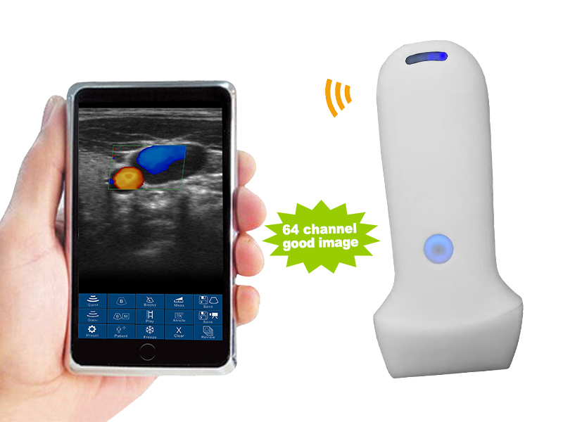
Suitable Handheld Wireless Color Doppler Ultrasound for Physical Medicine
Linear array


- Linear modes.
- Comprehensive connectivity with support for iOS, Android, and Windows platforms.
- Frequency: Linear probe 7.5MHz/10.0MHz
- Display mode: B, B/M, color doppler version with B+Color, B+PDI, B+PW
- Display Depth: Linear 20/40/60/80mm
- Measure: Length, Area, Velocity, HR, S/D, Auto Blood Vessel Measurement
- Weight: 180g
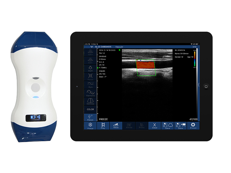
Double Headed Handheld Wireless Color Doppler Ultrasound for Physical Medicine
Linear array Frequency: 7.5MHz/10.0MHz & 10MHz/12.0MHz


- Linear modes.
- Comprehensive connectivity with support for iOS, Android, and Windows platforms.
- Frequency: Linear probe 7.5MHz/10.0MHz & 10MHz/12.0MHz
- Display mode: B, B/M, color doppler version with B+Color, B+PDI, B+PW
- Display Depth: Linear 20/40/60/80mm
- Measure: Length, Area, Velocity, HR, S/D, Auto Blood Vessel Measurement
- Weight: 250g
Key Applications of Handheld Wireless Color Doppler Ultrasound in Physical Medicine
Handheld wireless color Doppler ultrasound has become an essential tool in physical medicine due to its versatility, portability, and ability to provide real-time diagnostic imaging at the point of care. This technology is particularly useful for musculoskeletal, vascular, and nerve evaluations, and for guiding various therapeutic procedures.
Here are the key applications of handheld wireless color Doppler ultrasound in physical medicine, along with details on how it enhances patient care:
1. Musculoskeletal Injury Diagnosis
- Application: Diagnosis of muscle, tendon, and ligament injuries, common in sports medicine and rehabilitation.
- Details:
- Tendon Tears and Tendinopathies: Handheld ultrasound provides detailed imaging of tendon integrity and blood flow (via Doppler), enabling the diagnosis of partial or full-thickness tears in tendons such as the Achilles, rotator cuff, or patellar tendon.
- Ligament Injuries: Ultrasound can assess ligament damage, including ACL, MCL, and LCL injuries, offering real-time imaging to evaluate structural integrity and guide treatment.
- Muscle Strains and Tears: Physicians can identify the degree of muscle injury and hematoma formation, making it easier to create effective rehabilitation plans.
2. Guided Injections and Interventional Procedures
- Application: Assisting with precise joint injections, aspirations, and nerve blocks.
- Details:
- Joint Injections: Real-time ultrasound guidance ensures accurate placement of medications such as corticosteroids or hyaluronic acid into the joint space, improving outcomes in conditions like osteoarthritis.
- Nerve Blocks: In pain management, ultrasound guides nerve blocks, ensuring that anesthetics or anti-inflammatory agents are delivered precisely near the nerve, minimizing complications and improving efficacy.
- Bursa and Cyst Aspiration: For conditions like bursitis or Baker’s cysts, ultrasound-guided aspiration allows for the removal of excess fluid with minimal discomfort and improved accuracy.
3. Vascular Assessment and Monitoring
- Application: Evaluation of blood flow and vascular health in patients with conditions such as deep vein thrombosis (DVT), peripheral artery disease (PAD), and varicose veins.
- Details:
- Color Doppler for Blood Flow: Color Doppler imaging detects vascular abnormalities like venous insufficiency or arterial blockages, ensuring timely diagnosis and intervention in patients at risk of thrombosis or compromised circulation.
- DVT Diagnosis: Portable ultrasound can quickly assess for deep vein thrombosis, especially in post-operative or immobilized patients, reducing the risk of pulmonary embolism.
- Peripheral Artery Disease: Ultrasound helps monitor arterial stenosis or occlusions, providing clinicians with crucial data to manage rehabilitation in patients with PAD or those recovering from surgery.
4. Nerve Entrapment Syndromes
- Application: Diagnosing and managing nerve compression disorders such as carpal tunnel syndrome or cubital tunnel syndrome.
- Details:
- Nerve Compression Diagnosis: Handheld ultrasound can visualize nerve swelling and compression, offering a clear image of median nerve entrapment in the wrist or ulnar nerve entrapment in the elbow.
- Guided Therapy: Ultrasound can assist in guiding nerve release surgeries or injections of anti-inflammatory agents for nerve relief, improving patient outcomes in nerve entrapment syndromes.
5. Dynamic and Functional Musculoskeletal Imaging
- Application: Dynamic assessments of joints, tendons, and muscles during movement to identify functional impairments or biomechanical issues.
- Details:
- Joint Function Assessment: Ultrasound allows physicians to observe joint movement in real time, identifying issues such as joint instability, frozen shoulder, or rotator cuff impingement while the patient performs active motions.
- Tendon Gliding and Function: Dynamic imaging is used to assess tendon movement during stretching or exercise, helping to detect issues like tendon adhesions or partial tears that may affect motion and strength.
- Real-Time Feedback: This capability is crucial in rehabilitation, as it allows practitioners to monitor progress and adjust therapeutic exercises based on real-time functional imaging.
6. Soft Tissue Evaluation
- Application: Assessing soft tissue injuries, such as bursitis, fasciitis, and scar tissue formation, particularly in post-surgical recovery or chronic conditions.
- Details:
- Bursitis and Tendinitis: Ultrasound can detect inflammation of the bursae and tendons, guiding appropriate treatment plans including aspirations, injections, or targeted physical therapy.
- Scar Tissue Monitoring: Ultrasound helps evaluate fibrosis or scar tissue formation after surgery or trauma, aiding in developing rehabilitation programs to improve mobility and function.
- Myofascial Pain: It can visualize trigger points and areas of muscle stiffness, allowing for precise treatment using trigger point injections or myofascial release therapy.
7. Fracture Healing and Monitoring
- Application: Monitoring fracture healing and bone callus formation in patients recovering from injuries or surgeries.
- Details:
- Soft Tissue and Bone Monitoring: Although ultrasound is limited for bone imaging, it is highly effective in evaluating the healing of soft tissues around a fracture, detecting early signs of non-union or delayed healing.
- Post-Surgical Follow-Up: Ultrasound helps monitor the progress of healing in patients after fracture repairs, guiding rehabilitation and early intervention if complications arise.
8. Chronic Pain Management
- Application: Providing imaging for managing chronic pain conditions like myofascial pain syndrome, fibromyalgia, and chronic tendinitis.
- Details:
- Guided Pain Interventions: Ultrasound helps physicians target pain sources by guiding injections into trigger points, inflamed tendons, or swollen bursae, improving accuracy and reducing patient discomfort.
- Nerve Pain Management: Ultrasound guidance for peripheral nerve blocks ensures precise delivery of anesthetics or steroids for long-term pain relief in conditions like sciatica or neuropathic pain.
Handheld wireless color Doppler ultrasound is a powerful tool in physical medicine, offering real-time, non-invasive imaging that enhances diagnostic accuracy and therapeutic outcomes. From musculoskeletal assessments to vascular evaluations, it supports precise, point-of-care decision-making, improving patient care in rehabilitation, sports medicine, and chronic pain management. The versatility, portability, and wireless connectivity of these devices make them indispensable in delivering high-quality care in both clinical and remote settings.
Choosing the ideal Handheld Wireless Color Doppler Ultrasound for Physical Medicine: Key Factors to Consider
When selecting a handheld ultrasound device for physical medicine, it’s crucial to ensure that the device meets the specific needs of the practitioner and patients. Handheld ultrasound devices have transformed diagnostic and therapeutic approaches in musculoskeletal, vascular, and soft tissue evaluations. To choose the best option, here are the key factors to consider:
1. Portability and Ease of Use
Key Considerations:
- Size and Weight: The device should be lightweight and portable, making it easy to carry between different treatment rooms or facilities, especially for clinicians who work in remote areas or make home visits.
- Wireless Connectivity: Devices with wireless capability offer better mobility and reduce cable clutter, improving ease of use in busy clinical environments.
Why It Matters: In physical medicine, practitioners often need to move quickly between patients, whether in clinics or in-field settings. A lightweight, wireless handheld ultrasound allows for easy point-of-care diagnostics without having to transport bulky equipment.
2. Image Quality and Resolution
Key Considerations:
- High-Resolution Imaging: Look for devices that offer high-resolution 2D images and color Doppler capabilities to accurately visualize muscle, tendon, ligament, and vascular structures.
- Frequency Range: Ultrasound devices with a wider frequency range are ideal for examining both superficial and deeper tissues. For example, 7-10 MHz probes are better for detailed imaging of tendons, while lower frequency probes can assess deeper structures like joints or vessels.
Why It Matters: For musculoskeletal evaluations, clear and detailed imaging is essential to identify tendon tears, ligament damage, or vascular abnormalities. High image resolution ensures accurate diagnosis and helps guide effective treatments, such as guided injections or nerve blocks.
3. Durability and Battery Life
Key Considerations:
- Battery Longevity: A longer battery life ensures the device remains operational throughout the day without frequent recharging. Aim for a device that offers 4 hours of continuous use.
- Ruggedness: Look for devices that are shock-resistant and designed for clinical environments, where equipment might be exposed to drops, liquids, or other hazards.
Why It Matters: In fast-paced clinical or outpatient settings, handheld ultrasound devices need to withstand frequent use and last through long working hours. Durable devices with extended battery life are essential for uninterrupted patient care, particularly when used in multiple treatment sessions or during fieldwork.
4. Probe Versatility and Compatibility
Key Considerations:
- Multi-Frequency Probes: Some devices offer interchangeable probes for different applications (e.g., linear, curved, or phased-array probes) or multi-frequency probes that adjust to various depths.
- Specialized Probes: Certain probes are more effective for specific tasks—linear probes are ideal for superficial structures, while curved probes work better for deeper tissues, such as in abdominal or pelvic scans.
Why It Matters: In physical medicine, clinicians often need to switch between applications such as musculoskeletal imaging, vascular assessments, and guided interventions. Having a device with versatile probes ensures a wider range of diagnostic and therapeutic uses without needing multiple devices.
5. Doppler Functionality
Key Considerations:
- Color Doppler Capabilities: Devices with color Doppler functionality can visualize blood flow and vascular structures, which is crucial for assessing vascular conditions in physical medicine.
- Power Doppler: Power Doppler ultrasound can detect low-velocity blood flow, making it valuable for inflammation and chronic pain diagnoses where vascular involvement is suspected.
Why It Matters: Vascular assessments are essential in managing conditions like deep vein thrombosis (DVT), peripheral artery disease (PAD), or in monitoring post-surgical healing. Doppler ultrasound is indispensable in evaluating blood flow and identifying vascular abnormalities in rehabilitation settings.
6. Affordability and Cost-Effectiveness
Key Considerations:
- Cost: Handheld ultrasound devices vary widely in price depending on their features. It’s important to balance cost with the features that will be most useful in your practice.
- Return on Investment (ROI): Devices that offer multiple functionalities (e.g., musculoskeletal, vascular, and nerve imaging) provide greater value in the long run, as they reduce the need for multiple devices.
Why It Matters: Many physical medicine clinics operate on tight budgets. Choosing a cost-effective device that meets most diagnostic needs without sacrificing essential features ensures better patient care without financial strain.
7. Learning Curve and Support
Key Considerations:
- Ease of Use: The device should have an intuitive interface that is easy to learn for both experienced practitioners and those new to ultrasound technology.
- Training and Support: Supplier that offer Basic operational training programs, user manuals, and customer support can help clinicians quickly become proficient in using the device.
Why It Matters: Clinicians in physical medicine may not always be experts in using ultrasound technology. A device with a gentle learning curve and strong customer support ensures that users can take full advantage of the ultrasound’s capabilities without extensive delays or training.
3. FDA and CE Approval
- Significance: Regulatory approval ensures that the device meets the safety and performance standards required for medical use.
- What to look for: Ensure the device has FDA approval or CE marking, especially for key applications in internal medicine such as abdominal and lung imaging.
Choosing the ideal handheld ultrasound for physical medicine involves balancing image quality, portability, connectivity, and affordability. By considering factors such as real-time feedback, Doppler capabilities, and ease of use, practitioners can select a device that enhances diagnostic accuracy, improves patient outcomes, and streamlines workflows. A well-chosen ultrasound system will provide valuable support in evaluating musculoskeletal injuries, guiding injections, monitoring vascular health, and aiding rehabilitation efforts in clinical and field settings.
SonoWave Technologies
E-mail: info@sonowavetech.com
WhatsApp: +8801717 811 312
Address: 205/4, Begum Rokeya Sharani, Agargaon, Dhaka-1207, Bangladesh
