Handheld Wireless Color Doppler Ultrasound for
Cardiology.
Instant Cardiac Solutions at Your Fingertips
When selecting handheld ultrasound devices for cardiology, several factors such as image quality, ease of use, advanced features like Doppler imaging, and integration with other medical devices should be considered. Here are some of the best handheld ultrasound devices that are particularly suited for cardiology applications:
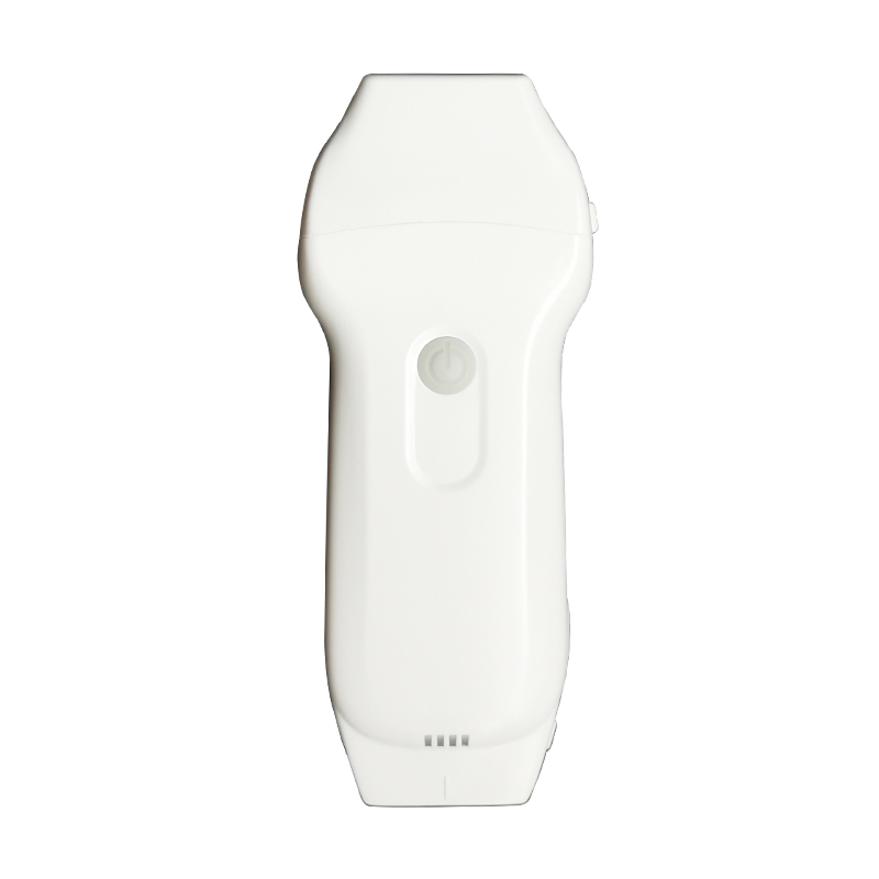
Handheld Wireless Color Doppler Ultrasound
Cardiac + Linear array


- 3-in-1 scanning with Cardiac and Linear modes.
- Comprehensive connectivity with support for iOS, Android, and Windows platforms.
- Frequency: Phased Probe 2.2/3.6MHz,Linear Probe 7.5/10MHz
- Display mode: B, B/M, color doppler version with B+Color, B+PDI, B+PW
- Display Depth: Phased 90~190mm, Linear 20~100mm
- Measure: Length, Area, Velocity, HR, S/D, LVIDd, LVIDs, EF, SV, Auto Vessel Measure (TAMEAN, Flow, Diam)
- Weight: 150g
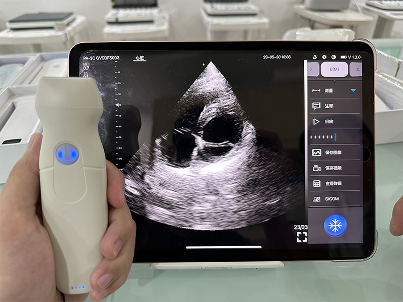
Handheld Wireless Color Doppler Ultrasound
Cardiac/Phased array


- Phased/Cardiac modes.
- Comprehensive connectivity with support for iOS, Android, and Windows platforms.
- Frequency: Central 2.8MHz, cardiac reverse harmonic 3.6mhz, and the transcranial fundamental 2.2MHz
- Display mode:B, B/M, and Color, PW, CW, PDI
- Display Depth: 90/160/200/240mm
- Measure: Length, Area, Angle, Heart, Velocity, HR, S/D, LVIDd, LVIDs, EF, SV
- Weight: 120g
Key Applications
Handheld wireless color doppler ultrasound devices offer cardiologists the ability to perform real-time imaging and assessments directly at the point of care. Here are the key applications of handheld wireless ultrasound for cardiologists:
1. Transthoracic Echocardiography (TTE)
- Application: Non-invasive evaluation of cardiac structure and function.
- Details: Assess ventricular size, wall motion, valve function, ejection fraction, and pericardial effusion. Handheld devices allow rapid bedside evaluation, making TTE more accessible for routine checks and emergent situations.
2. Heart Failure Management
- Application: Monitoring heart function in patients with heart failure.
- Details: Evaluate left ventricular function, detect fluid buildup, and adjust treatments such as diuretics based on the patient’s cardiac function. Wireless ultrasound enables frequent monitoring in both in-patient and out-patient settings.
3. Valvular Heart Disease Assessment
- Application: Evaluation of valve disorders like stenosis and regurgitation.
- Details: Handheld devices, particularly those with Doppler imaging, can assess blood flow across the heart valves, aiding in diagnosing conditions like mitral regurgitation, aortic stenosis, and tricuspid valve abnormalities.
4. Pericardial Effusion Detection
- Application: Identifying fluid around the heart.
- Details: Wireless ultrasound can quickly detect pericardial effusion and assess its severity, allowing cardiologists to intervene in cases of cardiac tamponade or other urgent conditions.
5. Cardiac Function Assessment in Acute Care
- Application: Immediate evaluation of cardiac function in emergency or critical care.
- Details: Handheld devices can be used in emergency rooms, intensive care units, or during cardiac arrests (Code Blue scenarios) to quickly assess heart function, including wall motion abnormalities, during resuscitation.
6. Ejection Fraction Measurement
- Application: Monitoring and measuring left ventricular ejection fraction (LVEF).
- Details: Assessing ejection fraction is critical in diagnosing and managing heart failure. Many handheld ultrasound devices feature AI-assisted tools for calculating LVEF quickly, improving the speed and accuracy of assessments.
7. Pulmonary Hypertension Diagnosis
- Application: Evaluating right heart function and pulmonary artery pressure.
- Details: Cardiac ultrasound can assess right ventricular function and estimate pulmonary artery pressure, helping diagnose and manage pulmonary hypertension, which often requires ongoing monitoring.
8. Arrhythmia and Cardiac Chamber Size Assessment
- Application: Identifying atrial or ventricular enlargement due to arrhythmias or structural abnormalities.
- Details: Cardiac ultrasound can evaluate the size of the atria and ventricles, providing insight into conditions like atrial fibrillation or hypertrophic cardiomyopathy.
9. Post-Intervention Follow-Up
- Application: Monitoring patients after cardiac interventions such as stent placement, valve repair, or bypass surgery.
- Details: Handheld ultrasound provides a convenient way to monitor recovery and detect complications early by visualizing heart function and detecting any signs of restenosis or valve failure.
10. Cardiac Arrest Evaluation (Peri-Resuscitation Imaging)
- Application: Guiding resuscitation efforts during cardiac arrest.
- Details: Handheld ultrasound can help assess the effectiveness of compressions, identify reversible causes of arrest (such as cardiac tamponade or massive pulmonary embolism), and evaluate heart function during resuscitation.
11. Congenital Heart Disease Monitoring
- Application: Ongoing assessment of congenital heart defects in children and adults.
- Details: Wireless ultrasound enables non-invasive, frequent monitoring of congenital heart defects like septal defects, coarctation of the aorta, and valve anomalies, especially in out-patient settings.
12. Myocardial Infarction (MI) Assessment
- Application: Rapid evaluation of myocardial infarction and associated complications.
- Details: Assessing regional wall motion abnormalities and ventricular function during or after an MI is critical. Handheld ultrasound provides a quick, portable solution to monitor heart function and detect complications such as aneurysm or rupture.
13. Guiding Cardiac Interventions
- Application: Assisting in procedures such as pericardiocentesis or transcatheter interventions.
- Details: Handheld ultrasound can be used to guide needle placement for pericardiocentesis or catheter placement in structural heart disease interventions, reducing the risk of complications.
14. Cardiomyopathy Diagnosis
- Application: Identifying and managing hypertrophic, dilated, or restrictive cardiomyopathy.
- Details: Cardiac ultrasound allows visualization of ventricular wall thickness, chamber size, and overall heart function, helping diagnose and monitor patients with cardiomyopathies.
15. Preoperative Cardiac Evaluation
- Application: Cardiac risk assessment before surgery.
- Details: Handheld ultrasound can be used to assess cardiac function in patients with underlying heart disease before undergoing non-cardiac surgery, reducing the risk of perioperative cardiac events.
16. Remote and Rural Cardiovascular Care
- Application: Delivering cardiology care in remote areas where access to traditional imaging equipment is limited.
- Details: Wireless handheld ultrasound allows cardiologists to perform cardiac assessments in rural or underserved areas, with the ability to share images and data remotely for consultation or follow-up.
17. Hypertension Management
- Application: Evaluating heart structure and function in hypertensive patients.
- Details: Long-standing hypertension can cause left ventricular hypertrophy (LVH) and other structural changes. Handheld ultrasound allows for regular monitoring of these changes in out-patient settings.
18. Pulmonary Embolism Diagnosis
- Application: Evaluating right heart strain in suspected pulmonary embolism cases.
- Details: Handheld ultrasound can be used to assess the right ventricular size and function, detecting signs of right heart strain that suggest a pulmonary embolism.
19. Stress Echocardiography
- Application: Monitoring heart function under stress.
- Details: Handheld ultrasound can be used to evaluate changes in heart function during or after physical or pharmacological stress tests, helping detect ischemia or functional abnormalities.
20. Aortic Disease Monitoring
- Application: Assessing the aorta for aneurysms or dissection.
- Details: Handheld ultrasound can detect dilatation of the ascending aorta or signs of aortic dissection, allowing cardiologists to intervene early.
Handheld wireless color doppler ultrasound devices provide cardiologists with a flexible, portable, and efficient tool for diagnosing, monitoring, and managing a wide range of cardiac conditions. From assessing heart function and diagnosing heart failure to monitoring post-intervention recovery, these devices offer real-time imaging and diagnostic capabilities that improve patient care both in hospital settings and remote locations.
Choosing the ideal Handheld Ultrasound for Cardiologists: Key Factors to Consider
Choosing the ideal handheld wireless color doppler ultrasound for cardiologists involves evaluating several important factors to ensure that the device meets the demands of cardiovascular care. Here are the key factors to consider:
1. Image Quality
- Importance: Cardiac imaging requires high-resolution images to assess heart function, structure, and blood flow accurately.
- Consideration: Ensure the device provides high-quality 2D imaging with clear visualization of cardiac structures such as chambers, valves, and vessel walls. Look for devices with advanced imaging technologies like harmonic imaging or speckle reduction.
2. Doppler Capabilities
- Importance: Doppler imaging is essential for evaluating blood flow through the heart, vessels, and valves, which is critical in diagnosing conditions like valvular stenosis, regurgitation, and heart failure.
- Consideration: Choose a handheld ultrasound that offers Color Doppler, Pulsed-wave Doppler, and Continuous-wave Doppler for comprehensive blood flow assessment.
3. Cardiac-Specific Presets
- Importance: Pre-configured cardiac presets optimize imaging for specific heart views (e.g., parasternal long-axis, apical, subcostal views) and streamline workflows.
- Consideration: Look for devices that include dedicated cardiac presets, as they ensure optimal settings for echocardiography and cardiac assessment.
4. Portability and Ergonomics
- Importance: Handheld ultrasound devices should be lightweight, portable, and easy to handle for quick bedside assessments and emergency use.
- Consideration: Select a device that is compact and easy to transport, especially if you’re using it in different settings (e.g., ICU, clinic, or remote locations). Wireless devices offer greater flexibility and ease of use.
5. Battery Life
- Importance: Cardiologists often need to perform multiple scans in one session, especially in busy settings like emergency departments.
- Consideration: Opt for a device with a long battery life, ideally lasting several hours of continuous scanning. Quick charging capabilities are also an advantage.
6. Ease of Use and Workflow Integration
- Importance: The device should have an intuitive interface, allowing fast and easy use for both routine and emergency cardiac evaluations.
- Consideration: Look for devices that provide user-friendly interfaces, easy navigation, and minimal setup time.
7. Wireless Connectivity and Data Sharing
- Importance: The ability to share images and results quickly with other medical professionals is crucial, particularly in telemedicine or remote consultations.
- Consideration: Devices with wireless connectivity, make it easier to upload, review, and share cardiac ultrasound images with the broader care team.
8. Durability and Water Resistance
- Importance: In emergency situations or field conditions, handheld devices may be exposed to fluids or rough handling.
- Consideration: Ensure the device is durable, shock-resistant, and ideally has some degree of water resistance to withstand varied clinical environments.
10. Price and Cost-Effectiveness
- Importance: The device should fit within your budget while delivering the necessary features for quality cardiac care.
- Consideration: Evaluate the cost of the handheld device, including additional fees for software updates, accessories, and warranties. Consider whether the features justify the investment in terms of time saved and clinical effectiveness.
11. Transducer Type and Versatility
- Importance: Cardiology requires specific transducer types, such as phased array transducers, for echocardiography.
- Consideration: Choose a device with a phased array probe, which is optimal for imaging deep structures like the heart. We offer multi-mode probes that switch between linear, curved, and phased array, adding versatility for broader applications beyond cardiology.
12. Software Updates and Support
- Importance: Regular software updates ensure the device remains functional and compliant with evolving healthcare standards.
- Consideration: Check for manufacturers that offer ongoing software updates, customer support, and training resources. Devices that improve over time with new software features provide long-term value.
3. FDA and CE Approval
- Importance: Regulatory approval ensures that the device meets safety and performance standards.
- Consideration: Ensure the handheld ultrasound device is FDA-approved (in the U.S.) or CE-marked (in Europe) for cardiac applications, ensuring it meets the regulatory standards for medical devices.
By considering these factors, cardiologists can select a handheld ultrasound device that fits their clinical needs, enhances workflow efficiency, and delivers high-quality cardiac care across various healthcare settings.
SonoWave Technologies
E-mail: info@sonowavetech.com
WhatsApp: +8801717 811 312
Address: 205/4, Begum Rokeya Sharani, Agargaon, Dhaka-1207, Bangladesh
