Handheld Wireless Color Doppler Ultrasound for Anesthesiologists
Handheld wireless color Doppler ultrasound technology is a crucial tool for anesthesiologists, offering real-time imaging and enhanced precision in a variety of applications. It is used for guiding peripheral nerve blocks, central venous catheterization, and arterial line insertion, improving the accuracy and safety of these procedures. The technology also aids in hemodynamic monitoring, cardiac assessments, and pulmonary embolism detection, providing real-time insights during surgeries and critical care. Its portability, ease of use, and ability to visualize blood flow make it indispensable for enhancing patient care in anesthesia.

Handheld Wireless Color Doppler Ultrasound for anesthesia
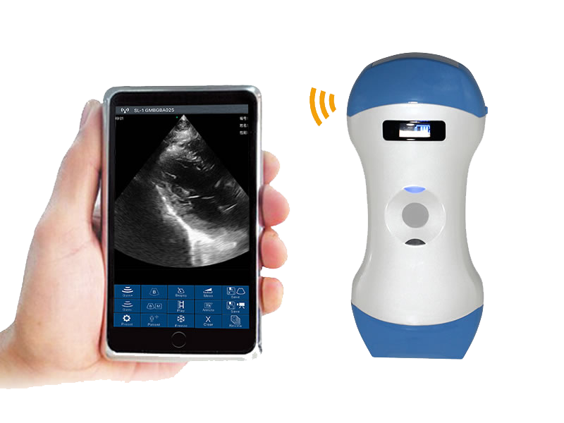
Convex/Phased + Linear array


- 3-in-1 scanning with Convex/Phased and Linear modes.
- Comprehensive connectivity with support for iOS, Android, and Windows platforms.
- Frequency: Convex/Phasedarray probe 3.2MHz/5MHz, Linear probe 7.5MHz/10.0MHz
- Display mode: B, B/M, color doppler version with B+Color, B+PDI, B+PW
- Display Depth: Convex 90/160/220/305mm, Linear 20/40/60/80mm
- Measure: Length, Area, Velocity, HR, S/D, GA (CRL, BPD, GS, FL, HC, AC), EFW (BPD, FL), LVIDd, LVIDs, EF, SV, Auto Vessel Measure (TAMEAN, Flow, Diam)
- Weight: 250g

Handheld Wireless Color Doppler Ultrasound for anesthesia
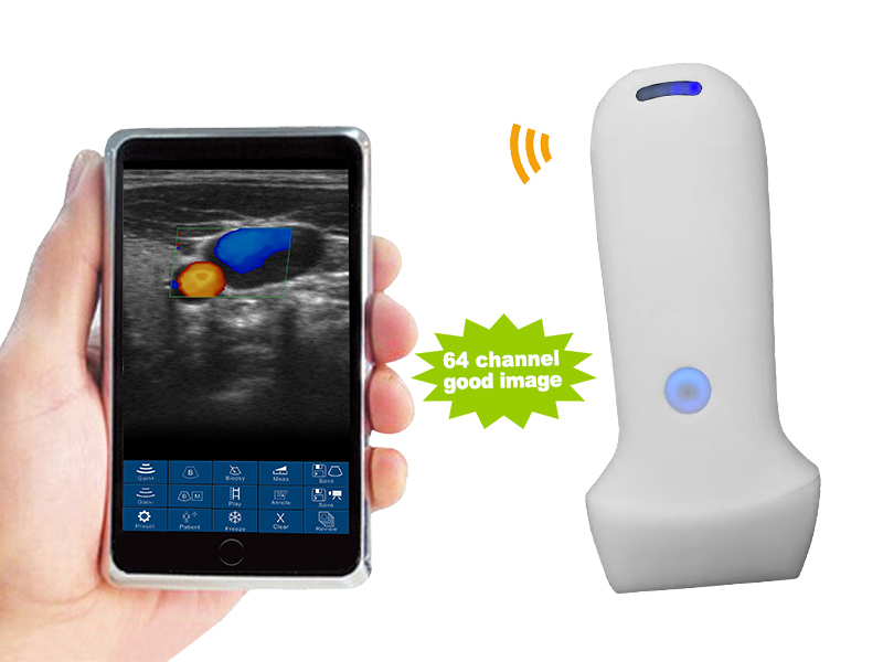
Linear array Frequency: 7.5MHz/10.0MHz


- Linear modes.
- Comprehensive connectivity with support for iOS, Android, and Windows platforms.
- Frequency: Linear probe 7.5MHz/10.0MHz
- Display mode: B, B/M, color doppler version with B+Color, B+PDI, B+PW
- Display Depth: Convex 90/160/220/305mm, Linear 20/40/60/80mm
- Measure: Length, Area, Velocity, HR, S/D, Auto Blood Vessel Measurement
- Weight: 180g
Key Applications of Handheld Wireless Color Doppler Ultrasound for Anesthesiologists
Handheld wireless color Doppler ultrasound technology has become an essential tool for anesthesiologists, particularly for improving accuracy, safety, and efficiency in various anesthesia-related procedures. With its portability and real-time imaging capabilities, this technology enhances patient care in a wide range of clinical settings. Below are the key applications of handheld wireless color Doppler ultrasound for anesthesiologists:
1. Peripheral Nerve Blocks
- Application: Used to guide the placement of needles for regional anesthesia or nerve blocks.
- Details:
- Real-Time Visualization: The ultrasound allows anesthesiologists to visualize nerves, surrounding structures, and the needle pathway.
- Color Doppler: Helps avoid puncturing blood vessels by identifying arterial and venous flow near the nerve, reducing the risk of complications like hematomas.
2. Central Venous Catheterization
- Application: Assisting in the placement of central venous catheters in critically ill patients or during surgeries.
- Details:
- Vein Identification: Handheld color Doppler ultrasound is used to locate the internal jugular vein, subclavian vein, or femoral vein for accurate catheter insertion.
- Reduced Complications: Ultrasound guidance reduces the risk of arterial puncture, pneumothorax, and other catheter-related complications.
3. Arterial Line Insertion
- Application: Assisting in the insertion of arterial lines for hemodynamic monitoring during surgeries or in critical care.
- Details:
- Doppler Guidance: Color Doppler provides a clear view of arterial flow, allowing precise placement of arterial lines in patients with challenging vascular anatomy or poor perfusion.
- Real-Time Imaging: Minimizes failed attempts and reduces complications such as pseudoaneurysms.
4. Transesophageal Echocardiography (TEE) for Cardiac Anesthesia
- Application: Used during cardiac surgeries or for intraoperative cardiac assessment, TEE is crucial for monitoring heart function.
- Details:
- Color Doppler: Provides real-time visualization of blood flow, valve function, and cardiac output during anesthesia.
- Cardiac Function Assessment: Detects abnormalities such as valve dysfunction or myocardial ischemia in real-time, enabling anesthesiologists to adjust anesthesia and medications promptly.
5. Pulmonary Embolism Detection
- Application: Assisting in the diagnosis of pulmonary embolism (PE) in patients presenting with sudden onset respiratory distress or hypoxia.
- Details:
- Color Doppler Imaging: Helps detect thrombi in the pulmonary arteries and assess blood flow to the lungs.
- Real-Time Detection: Anesthesiologists can quickly identify a PE in emergency situations and initiate appropriate interventions.
6. Echocardiography for Hemodynamic Monitoring
- Application: Used for monitoring cardiac output, stroke volume, and other hemodynamic parameters during surgeries and critical care.
- Details:
- Portable Ultrasound: Handheld color Doppler devices provide real-time insights into cardiac function, fluid responsiveness, and preload during major surgeries.
- Immediate Feedback: Enables anesthesiologists to make quick, informed decisions about fluid management and the use of inotropes or vasopressors.
7. Airway Management
- Application: Ultrasound-guided assessment of the airway for predicting difficult intubations and guiding tracheostomy procedures.
- Details:
- Color Doppler: Used to identify vascular structures near the trachea or cricothyroid membrane, ensuring a safe pathway during emergency airway management.
- Anatomical Assessment: Ultrasound helps evaluate airway structures, reducing the risk of failed intubation or tracheal injuries.
8. Evaluation of Diaphragmatic Function
- Application: Assessing diaphragmatic movement and function, particularly in post-operative or ventilated patients.
- Details:
- Color Doppler: Helps assess the movement of the diaphragm and blood flow in the surrounding areas to evaluate for conditions like diaphragmatic paralysis or dysfunction.
- Real-Time Monitoring: This is especially useful in patients who have undergone thoracic surgery or those on long-term mechanical ventilation.
9. Guidance for Paracentesis and Thoracentesis
- Application: Assisting in the placement of needles for procedures like paracentesis (removal of fluid from the abdomen) and thoracentesis (removal of fluid from the pleural space).
- Details:
- Doppler Imaging: Identifies safe insertion points while avoiding vascular structures, improving the safety and success rate of fluid drainage procedures.
- Guided Drainage: Ensures proper needle placement, minimizing risks of perforating organs or causing vascular injury.
10. Regional Anesthesia in Pediatrics
- Application: Assisting in administering regional anesthesia for pediatric patients, where anatomical landmarks may be less defined.
- Details:
- Real-Time Visualization: Provides a clear view of pediatric anatomy, improving accuracy in needle placement for nerve blocks or vascular access.
- Color Doppler: Helps avoid critical structures like arteries in smaller, more delicate pediatric patients.
Handheld wireless color Doppler ultrasound is a game-changer for anesthesiologists, enhancing precision and safety across a range of applications, from nerve blocks and vascular access to cardiac and respiratory assessments. Its portability and real-time imaging capabilities make it an indispensable tool in both routine and emergency anesthesia care, improving patient outcomes and reducing procedural complications.
Choosing the ideal Handheld Wireless Color Doppler Ultrasound for Anesthesiologists: Key Factors to Consider
Selecting the right handheld wireless color Doppler ultrasound for anesthesiologists is essential for improving procedural accuracy, safety, and efficiency. With a wide range of applications in anesthesia, including nerve blocks, vascular access, and cardiac monitoring, it’s important to consider the following key factors when choosing the ideal device:
1. Imaging Quality and Resolution
- Why It Matters: Anesthesiologists need high-resolution imaging to clearly visualize nerves, blood vessels, and other critical structures during procedures like nerve blocks or catheter placement.
- What to Look For: Devices with excellent B-mode resolution for anatomical details and sensitive color Doppler for blood flow visualization, ensuring precise guidance during invasive procedures.
2. Portability and Battery Life
- Why It Matters: In fast-paced and mobile environments like the operating room or critical care settings, portability is key.
- What to Look For: Lightweight, compact devices with long-lasting battery life allow for flexibility in multiple clinical settings without frequent recharging or bulky equipment.
3. Doppler Capabilities
- Why It Matters: Color Doppler is essential for visualizing blood flow, which is crucial for guiding vascular access and assessing hemodynamics.
- What to Look For: Ensure the ultrasound has color Doppler, power Doppler, and spectral Doppler to accurately assess blood flow, vessel patency, and velocity measurements during procedures like central venous catheterization.
4. Probe Versatility and Frequency Range
- Why It Matters: Anesthesiologists work with a variety of patients, from pediatrics to adults, requiring versatile probe options for different depths and structures.
- What to Look For: A wide range of transducer options, including high-frequency linear probes for superficial structures and lower-frequency curvilinear or phased-array probes for deeper tissues, ensures versatility in applications such as nerve blocks, airway management, and cardiac assessments.
5. Durability and Infection Control
- Why It Matters: In anesthesia, equipment must endure frequent use, sterilization, and handling in sterile environments.
- What to Look For: Devices with durable, rugged designs and IP-rated for water and dust resistance. The device should be compatible with standard disinfection and sterilization protocols to ensure patient safety.
6. Cost and Value
- Why It Matters: While functionality is important, cost plays a significant role, especially in budget-conscious healthcare environments.
- What to Look For: Consider devices that provide a balance of necessary features and affordability. Focus on long-term value, including software updates, warranty, and customer support, rather than just upfront cost.
Clinical Support and Training
- Why It Matters: Anesthesiologists need ongoing support to stay current with advancements in ultrasound technology and techniques.
- What to Look For: Devices that come with comprehensive clinical education, online tutorials, and responsive technical support are beneficial for ensuring that anesthesiologists are fully equipped to use the device effectively.
3. FDA and CE Approval
- Significance: Regulatory approval ensures that the device meets the safety and performance standards required for medical use.
- What to look for: Ensure the device has FDA approval or CE marking, especially for key applications in internal medicine such as abdominal and lung imaging.
Choosing the right handheld wireless color Doppler ultrasound for anesthesiologists involves balancing high-resolution imaging, ease of use, and portability. Key considerations include Doppler capabilities for blood flow assessment, versatile probe options, and seamless integration into clinical workflows. Durability, infection control, and cost-effectiveness also play crucial roles in making the ideal choice, ensuring anesthesiologists can provide safe, precise care across diverse anesthesia applications.
SonoWave Technologies
E-mail: info@sonowavetech.com
WhatsApp: +8801717 811 312
Address: 205/4, Begum Rokeya Sharani, Agargaon, Dhaka-1207, Bangladesh
